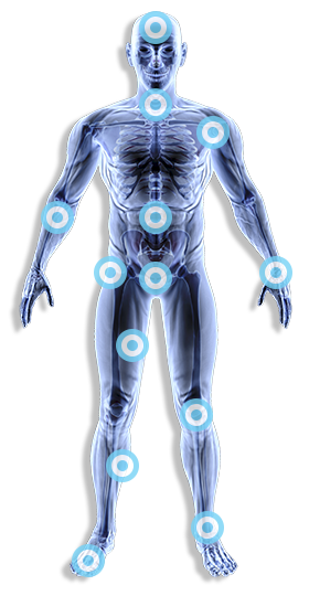
Aromioclavicular Joint / AC Joint
The AC joint is located at the tip of the shoulder where the acromion portion of the shoulder blade (scapula) and collarbone (clavicle) join together. The AC joint is not as mobile as the large main shoulder joint and only moves when the shoulder is overhead or across the chest (adducted).
The joint is partly filled with a thick pad of cartilage, known as the meniscus, which allows the joint to move. The AC joint is stabilised by its capsule and additional ligaments (coraco-clavicular ligaments).
What Can Cause Pain?
Inflammation due to over use or injury occurs mainly in adults over 30, it can be related to heavy lifting and bodybuilding. People with AC joint pain usually point with one finger to the pain being on top of the joint. The pain occurs on overhead movements and movement across the chest. The joint capsule and ligaments can be pain generators.
Osteoarthritis cause similar pain but there may not be any injury or precipitating factors. Osteoarthritis of the AC joint makes the joint more prominent and bigger. This bigger joint may rub against the rotator cuff tendons as they pass under the joint and cause pain similar to impingement over the outer aspect of the shoulder. This condition usually occurs in the 4-5 decade.
High impact injury can disrupt the joint. We see injuries as a consequence of rugby, football or hockey. Ligaments can be partially torn or ruptured. Various research papers and text books grade these injuries 1-6. Grade 1-2 being partial disruption to the ligaments around the joint. 3-6 involve complete severance of the ligaments around the joint.
Dependant on position, this may require surgical intervention, however appropriate rehabilitation might be the way forward.
Symptoms
Inflammation the joint might feel hot and swollen tender to touch, worse on lifting the arm across the body, compressive movement may also feel uncomfortable. Osteoarthritis, again pain could be fairly persistent but this tend to be more towards the end of full movement above your head, you are fairly specific in where the pain is, a common sign is the “finger sign” you point to that area of problem.
High impact injury Basically you know you have done something prior to the pain, sometime in high injury you can hear a pop.
What are some of the treatment can you have for the acromio-clavicular joint?
- Rest – a sling may be used initially after the injury.
- You may require pain killer over the counter or prescribed by your doctor.
- Cold therapy.
- Strengthening exercises for the shoulder.
- Specific active exercise program
- Muscle correction of imbalance
- Electrotherapy
- Ultrasound
- Frictional massage
- Injection therapy (OA if deemed appropriate)
Osteoarthritis is more commonly found in the fourth and fifth decade of life. It can be associated with previous trauma, repeated overhead work and play. This can sometimes respond to any of the above treatments however this joint does sometimes respond to injection if it is none responsive to conventional treatment with Ostenil (HA) usually up to 3 injections.
Broken Collar Bone / Clavical
The clavicle (collarbone) is the prominent bone on either side at the front of your shoulders and top of chest. The clavicle is the only bony link between the shoulder and the body itself, as well as providing protection to important underlying blood vessels and nerves. See: www.shoulderdoc.co.uk
A fractured collar bone is a crack or splintering of the collar bone (the bone that runs along the front of the shoulder to the breast bone, located just below your neck).
We see a lot of sportsmen and women in the clinic post injury. Some with severe traumas have required surgery reduction and fixation, others heal conservatively. If not managed correctly they can lead to further problems later.
Of course we also see also see the none sporting amongst you, again it is essential that the treatment and advice is the right one. Often people are discharge form A+E with a sheet of advice with little follow up, we see the secondary problems that arise from this.
What treatment is there for a fractured collar bone?
There are few things you can do yourself to ease the pain or heal the fracture. Treatment mainly involves resting the affected area in a sling in the initial phases. There are various types of slings available, if this has not been supplied we can help and advise.
It is very unusual for a collar bone fracture to require surgery, we see these in our Rugby league players, martial arts or high contact sports, however the elderly can be susceptible to displaced fractures due to there poor bone density.
A medical professional will also be able to provide interventions to help ease the pain such as appropriate medication. We have the necessary Physiotherapeutic equipment to compliment that during the healing phase. A fractured collar bone will take between four to eight weeks to heal.
Advice on types of activity will be provided throughout your rehabilitation. Recovery is usually complete with appropriate advice and treatment, the type of activities you are involved with will determine how and when to start. You may notice a bump where the fracture was for a few months after, or longer, but this shouldn’t cause any problems or pain.
Frozen Shoulder
Frozen shoulder often occurs with no explanation. It can present very suddenly (you can just wake up with this type of shoulder) some people may develop a frozen shoulder after a traumatic injury, but this is not always the case.
It restricts the movement in the shoulder; quite severely in some circumstances. Statistically diabetics and those with under active thyroids tend to be afflicted more than none diabetics, (endocrine disorders) and women are more likely then men to get this type of complaint.
The lining of the shoulder joint, known as the “capsule”, is normally a very flexible elastic structure. It’s looseness and elasticity allows the huge range of motion that the shoulder has. With a frozen shoulder this capsule (and its ligaments) becomes inflamed, swollen, red and contracted. The normal elasticity is lost and pain and stiffness set in.
There are some factors that make suffering from a frozen shoulder more common.
These include:
Shoulder trauma or surgery – if you have a shoulder injury frozen shoulder may occur, but not always. Surgery also increases the risk, especially if it is followed by a sustained period of joint immobilisation.
Your age range also statistically makes you have a higher risk of developing a frozen shoulder between 40-60 years of age.
Other systemic conditions – heart disease and Parkinson’s diseases have been associated with an increased risk of developing a frozen shoulder.
Patients with scar tissue in their hands, a condition called Dupuytrens contracture.
Phases of a frozen shoulder?
Three Phases
- Freezing
- Frozen
- Thawing
What are the symptoms of a frozen shoulder?
Pain and stiffness in the shoulder joint, pain at night when lying on the affected side and limited range of motion.
A frozen shoulder tends to have three “stages” to it:
You will suffer some bad pain particularly at night, with limited movement
The pain will ease off, but movement will become very limited sometimes as much as 50-60% its normal
Finally the shoulder loosens up and returns to normal with full movement.
It can take between two – three years for all these phases to occur. In younger people who are active, the whole process can sometimes be reduced to as little as 6 – 10 weeks. Injection therapy where appropriate might reduce that time.
Physiotherapy treatment
A frozen shoulder can be diagnosed on examination by our physiotherapists. An X-Ray was originally thought to be of no diagnostic value, however recent studies have suggested that this might form part of the overall management of this condition. X-ray can also confirm that there are no other problems or possible causes for the shoulder pain. See: www.shoulderdoc.com
Phase one. In the acute phase we have found that early cortisone injection has a good overall outcome to this condition. This should never be done in isolation of other treatment modalities. If thought necessary we can write to your GP to advice on appropriate drugs to be used and if required we can administer these at out clinic. Some people however will be unable to be administered an injection due to it being medically contraindicated.
During Phase two and three appropriate soft tissue and joint mobilisation is essential along with a tailored home exercise program. An injection can still be most useful at this point to accompany physiotherapy and home exercise.
Rotator Cuff Injury / Degeneration / Impingement
The shoulder comprises of 30 muscles. The rotator cuff muscles are:
- Supraspinatus
- Infraspinatus
- Teres Minor
- Subscapularis
The rotator cuff muscles predominantly stabilise the glenohumeral (shoulder joint), but also contribute significantly to movement. Any disruption of these muscles whether that is trauma, wear and tear, poor muscle imbalance or disease will have a significant bearing on how the shoulder works
A torn rotator cuff can happen following a trauma to the shoulder or just through “wear and tear”. Many athletes, like cricket players, swimmers, and javelin throwers, suffer of such injuries due to the repetitive movement of their shoulders.
Older people can develop degeneration in the rotator cuff associated with other problems such as arthritis in the shoulder.
Symptoms can sometimes vary, ranging from pain and loss of function, sometimes one or all muscles can be injured, called a “Massive cuff tear”
Sleeping on your arm may become difficult, combing your hair, or simply lifting a cup of tea or coffee can hurt. The pain is usually more pronounced when lifting up.
Later patients who have a rupture can be without pain but they find they need the assistance of the other arm to help lift it up, a massive tear is often seen with the “drop sign”, the person is unable to kept his arm elevated due to ruptured tendon.
Shoulder Dislocation
A dislocated shoulder is an injury in which your upper arm bone pops out of the cup-shaped socket that’s part of your shoulder blade. A dislocated shoulder is a more extensive injury than a separated shoulder, which involves damage to ligaments of the joint where the top of your shoulder blade meets the end of your collarbone.
The dislocation can be either front (anterior) and back (posterior) the most common is anterior. We tend to see people after injury and attendance at the local A+E department days or weeks later.
Other associated injuries that can occur with this type of trauma are sometimes not spotted or noted initially, we tend to see problems later on in our clinic or when referred by surgeons or specialists if not attending our clinic first.
Symptoms
They can vary from mild discomfort a few weeks after the injury to a great deal of discomfort and loss of function; this is usually because of a soliloquy of poor initial diagnostics or management.
What treatment can be done for shoulder dislocation?
Doctors treat dislocated shoulders by manipulating the shoulder back into place. This is usually followed up with an x-ray to ensure the shoulder has been put back into place properly, and no other bones were fractured around the shoulder as a result of the manipulation.
The arm is usually put in a sling and you should not use your arm for several weeks or until your consultant or Physiotherapist advices otherwise.
A rehabilitation program to exercise and restore the range of motion to the shoulder and strengthen the muscles is integral. They will also help to prevent future dislocations. Very few go on to re-dislocate if the appropriate management is given initially. Re-dislocation is higher in those patients under 20 years.
It is important to keep the range of movements in the elbow wrist and hand therefore gentle mobilisation of these areas should happen, progressing to small extension, flexion and rotation of the shoulder, depending on how painful the shoulder is. Certain movements should be avoided in the early phase of management.
After the shoulder has been rested you can enhance mobilisation and strength in that area through physiotherapy. If the shoulder is none responsive to this a labral tear must be excluded with MRI arthrogram , this is a special test to see if any of the soft tissues in your shoulder has been damaged.
Physiotherapists at the Physio Team-Works clinic have many years experience in dealing with these types of injuries, particularly in sport. Without the right care your recovery could be delayed. It is imperative that you get the right advice following this injury, otherwise your outcome will be less than successful.
Surgery
Success of neck surgery depends a lot on what happens in the pre and postoperative stages. It is important to maximise the surgery by doing exercises that help to stabilise, mobilise and protect the area. Your consultant may also recommend you have some sort of pre-operative intervention, we are ideally suited. This type of injury must not be left to its own devises.
Investigations may take the form of X–ray or MRI arthrogram. This is to identify whether certain tissue is damaged. Dependant on this is the type of surgery required.
Treatment
Dependent on the presentation, is it torn? Is it inflamed? Is it weak? Are the muscles not working as they should (poor patterning)? Thorough examination determines the course of management.
Most of the time, treatment for rotator cuff injuries involves exercise therapy. Our physiotherapists will design specific exercises to help heal your injury, improve the flexibility of your rotator cuff and shoulder muscles, and provide balanced shoulder muscle strength. Depending on the severity of your injury, physical therapy may take from several weeks to several months to reach maximum effectiveness.
Some of the treatment principles and modalities we employ in our clinic are as follows:
- Ultrasound
- Laser therapy
- Cryotherapy
- Thermotherapy
- Mobilizations
- Deep tissue massage techniques
- Specific graded exercise program
- Sport specific rehabilitation
- Bio-feedback machines
Surgery
Success of shoulder surgery depends a lot on what happens in the pre and postoperative stages. It is important to maximise the surgery by doing exercises that help to stabilise, mobilise and protect the area. Your consultant may also recommend you have some sort of pre-operative intervention, we are ideally suited.
Physio Team-Works will be able to guide you through these stages of rehabilitation. We can assist in monitoring your progress, setting your goals, and providing appropriate treatment to maximise your recovery potential. We can also inform you of how you can help your own recovery, and what should be avoided. You will be provided a specific rehabilitation program, and we aim to get you back to your full levels of activity and/or sport as quickly as possible.
Contact us
Physio Team-Works will be able to guide you through these stages of rehabilitation. We can assist in monitoring your progress, setting your goals, and providing appropriate treatment to maximise your recovery potential.
We can also inform you of how you can help your own recovery, and what should be avoided. You will be provided a specific rehabilitation programme, and we aim to back to your full levels of activity and/or sport as quickly as possible.
Call 01457 837 211 or complete our quick online form to arrange an appointment.


